Tendon overuse (Tendinopathy)
|
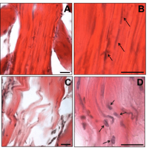
Histology of rat tendons(A & B), normal tendon, arrows indicate normal tendon cells with a spindle-like morphology; (C & D), overused tendon, arrows indicate rounded tendon cells. Scale bar = 25 μm. |
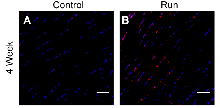
Higher Cathepsin L staining (red) at the insertion was found in the overuse group vs. control at 4 weeks. Scale bar = 100. μm (Seto, et al., Ann Biomed Eng. 2015 DOI 10.1007/s10439-014-1245-8). |
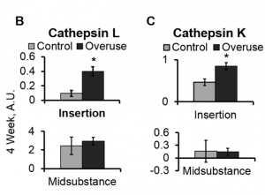
Cathepsins L (B) and K (C) activity at the insertion region after 4 weeks of overuse compared to age-matched controls. * indicates significantly greater activity over controls at the same timepoint (p<0.05) (Seto, et al., Ann Biomed Eng. 2015 DOI 10.1007/s10439-014-1245-8). |
|
||
Repair:
Cell delivery into tendon defects
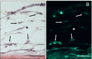 Hematoxylin & eosin staining (A) and fluorescent images (B) of tendon defects with mesenchymal stem cells (MSCs) encapsulated in hydrolytically degradable gels and cultured for 2 weeks.* indicates tendon tissue and arrows indicate encapsulated MSCs. Scale bar = 100 μm (Qiu, et al., Acta Biomater. 2010 DOI 10.1016/j.actbio.2010.11.002). |
PEG-based hydrogels |