Rotator Cuff Reattachment and Treatment
|
Images of supraspinatus muscle sections 1 (3+1) and 3 (3+3) weeks after surgical reattachment with control saline or therapeutic amnion treatments showing muscle fiber outlines (green) and regenerating fibers (red dots within fibers), quantified in graph. Amnion provides an improved early regenerative response. The Scale bar = 100um. (Anderson et al., Annals of Biomed Eng. 2021. 49: 3698-3710) |
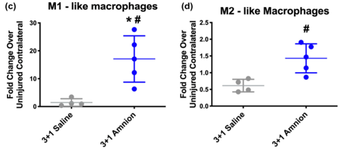
Quantification of macrophages 1 week after tendon reattachment after saline control or therapeutic amnion treatment. Early macrophage increase corresponds to the early regenerating fibers in amnion treated muscles. (Anderson et al., Annals of Biomed Eng. 2021. 49: 3698-3710) |
- Rotator cuff tears often require surgical intervention to reattach the torn tendon.
- Therefore, our lab has developed a model to assess therapeutic outcomes after surgical reattachment.
- In the photo and graph on the left, there is an increased number of regenerating fibers with therapeutic delivery after tendon reattachment.
- Early immune response can be evaluated and showed an increase with treatment after surgical reattachment (graphs on right).
- Our laboratory is working on delivery platforms to continue improving early outcomes in muscle after surgical reattachment of rotator cuffs.
- Click link below to read more from this paper on PubMed:
Reattachment and Treatment (Anderson et al)
Hydrogel Systems to Improve Mesenchymal Stem Cell Therapies and Delivery
|
|
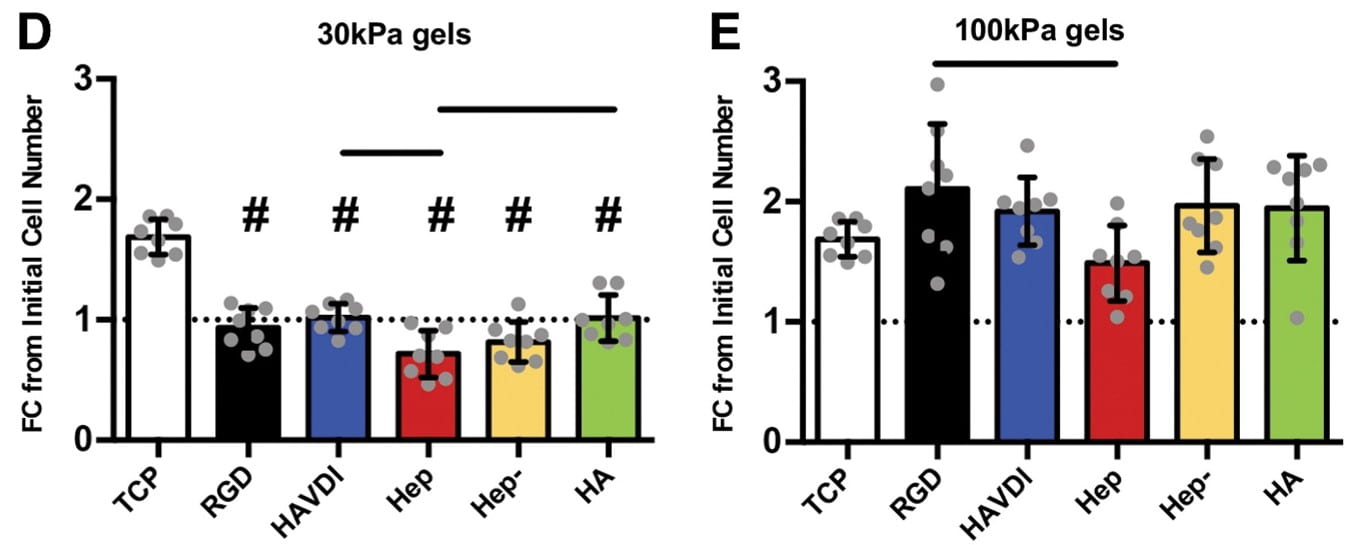
Different compressive stiffnesses (D) 30kPa and (E) 100kPa affect cell number after 4 days. (Ogle et al., Tiss Eng A. 2020. 26:1259-1271) |
Different compressive stiffnesses change the secretory capacity of mesenchymal stem cells with increases (red color) shown in the 30kPa hydrogels. (Ogle et al., Tiss Eng A. 2020. 26:1259-1271) |
- Expansion of mesenchymal stem cells using cell culture is necessary in order to reach quantities needed for therapeutic use.
- Therefore, the ability to maintain the therapeutic potency of mesenchymal stem cells during cell culture expansion is essential for tissue engineering applications.
- To address this, our lab is exploring hydrogel substrates that promote expansion and maintenance of therapeutic potential during cell culture.
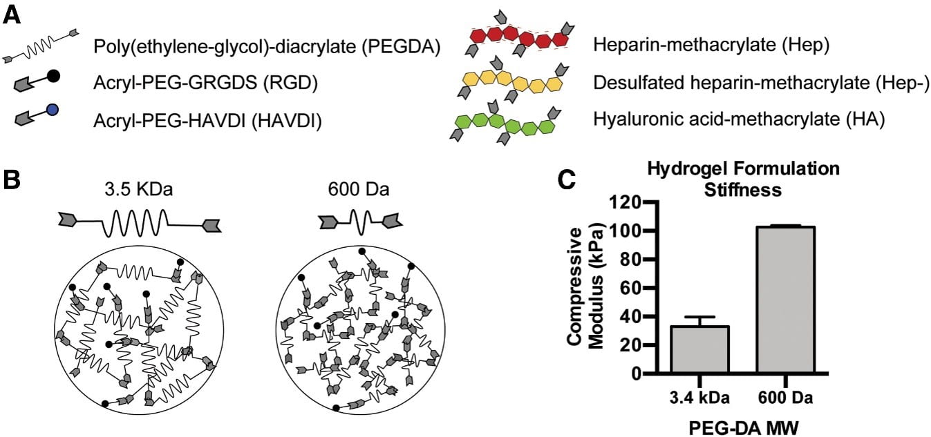
- The figure on the left shows that these hydrogel systems can vary in stiffness and biochemical composition.
- The different hydrogel formulations affect cell number (middle figure), secretory capacity (right figure) and senescence during culture.
- Our lab is working on utilizing these hydrogel systems for expansion and delivery of mesenchymal stem cells to treat rotator cuff muscle.
- Click link below to read more from this paper on PubMed:
Hydrogel Systems Stem Cells (Ogle et al)
Growth Factor Delivery after Rotator Cuff Tear
|
|
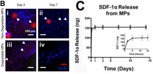
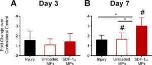
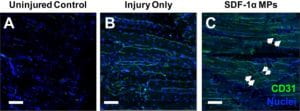
Quantification of mesenchymal stem cells at 3 and 7 days after rotator cuff injury in injured control muscles and muscles injected with MPs or SDF-1a loaded MPs. (Tellier et al., Regen Eng Trans Med. 2018. 4(2): 92-103) |
|
- After rotator cuff tendon tears, rotator cuff muscle damage is observed, including atrophy and fatty infiltration.
- In the photo on the left, there appears to be similar fibrous infiltration (blue staining) within the muscle (red) following injury compared to uninjured controls after one week.
- In the photo in the middle, the fibrous infiltration appears increased six weeks after injury in the injured muscle compared to the uninjured controls.
- Proteases such as metalloproteinases (MMPs) and cathepsins play a role in matrix damage after injury and are increased in injured muscles after rotator cuff tear (graphs on right).
- Our laboratory is working on means to treat muscle after rotator cuff tears.
- Click link below to read more from this paper on PubMed:
Growth Factor Delivery (Tellier et al)

